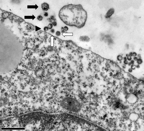
Figure 5.

Electron micrograph of HCV and HIV-1 extracellular particles in the vicinity of a K7 cell. Representative HIV-1 particles are indicated with black arrows, HCV with white arrows, and immature HIV-1 particles by an arrowhead. The HIV-1 particles are a little larger than HCV.
