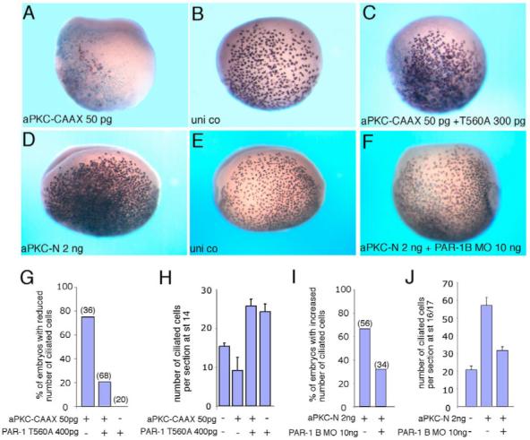
Fig. 5. aPKC functions upstream of PAR1 to specify ectodermal cell fates.

Embryos were injected with RNAs or MO as described in Fig. 1. Ciliated cells were detected at stages 14-16 by in situ hybridization with the α-tubulin probe. (A-C) T560A reverses the inhibitory effect of aPKC-CAAX on ciliated cell differentiation. Two sides of the same embryo are shown in B and C. (D-F) PAR1B MO (F) suppresses aPKC-N-mediated expansion of ciliated cells. The injected and uninjected sides of the same embryo are shown in D and E, respectively. (G-J) Quantification of the data shown in A-C (G,H) and D-F (I,J). Numbers of embryos per group are shown above bars. (G,I) Frequencies of embryos showing visible phenotypic changes. (H,J) Mean numbers of α-tubulin-positive cells per section±s.d. are shown. Sections of at least three representative embryos per group were analyzed.
