
Fig. 2. Embryos targeted for gata5 and gata6 fail to express GFP in a cmlc2:gfp reporter line.
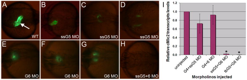
(A) A typical cmlc2:gfp embryo at 30 hpf shows GFP+ cardiomyocytes forming a heart tube (arrow). (B–G) Examples are shown representing cardiac phenotypes generated by injection of the ssG5 MO (B–D) and G6 MO (E–G). Co-injection of ssG5 MO and G6 MO results in an absence of GFP+ cardiomyocytes (H). Brightness of GFP appears lower in the reproduced images of B–G compared to A, because the injected embryos are slightly delayed, but also because the signal is more diffuse in the defective heart tubes. I: Quantitative real time PCR for cmlc2 transcripts. Endogenous cmlc2 transcript levels are reduced to approximately 1% the normal level in ssG5+6 and tbG5+6 double morphants (p<0.01).