
Fig. 6. The defect in gata5+6 morphants is specific to cardiac mesoderm.
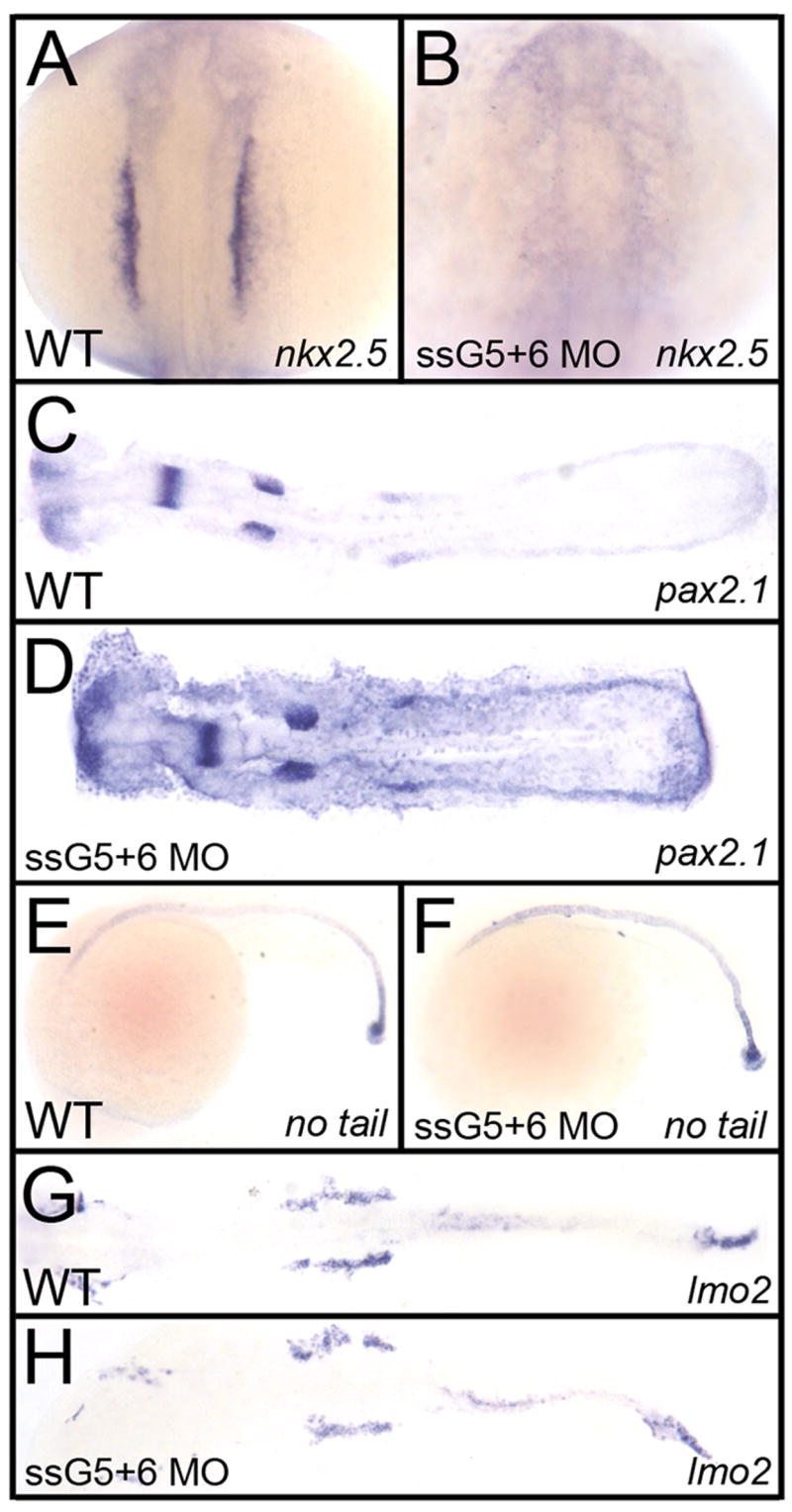
Shown in each panel is a typical representative embryo following processing by whole mount in situ hybridization. Embryos were either wild-type (A, C, E, G) or gata5 (ssG5) +6 double morphants (B, D, F, H). (A, B) The cardiomyocyte progenitor marker nkx2.5 at the 12 somite stage, shows the lack of cardiac progenitors in gata5+6 morphants. (C,D) The pronephric marker pax2.1 shows that the intermediate mesoderm was not affected in morphants. (E,F) The axial mesoderm marker no tail shows that gata5+6 morphants develop a normal notochord. (G, H) The lateral plate mesoderm, marked by the lmo2 probe, is also not altered in gata5+6 morphants. A,B: views are dorsal, with anterior to the top. C,D,G,H: embryos were flat mounted, views are dorsal, with anterior to the left. E,F: Views are from the left side, with anterior to the left. These panels represent patterns seen in A: 39/39; B: 38/38; C: 10/10; D: 23/23; E: 26/26; F: 45/45; G: 28/28; H: 42/42.