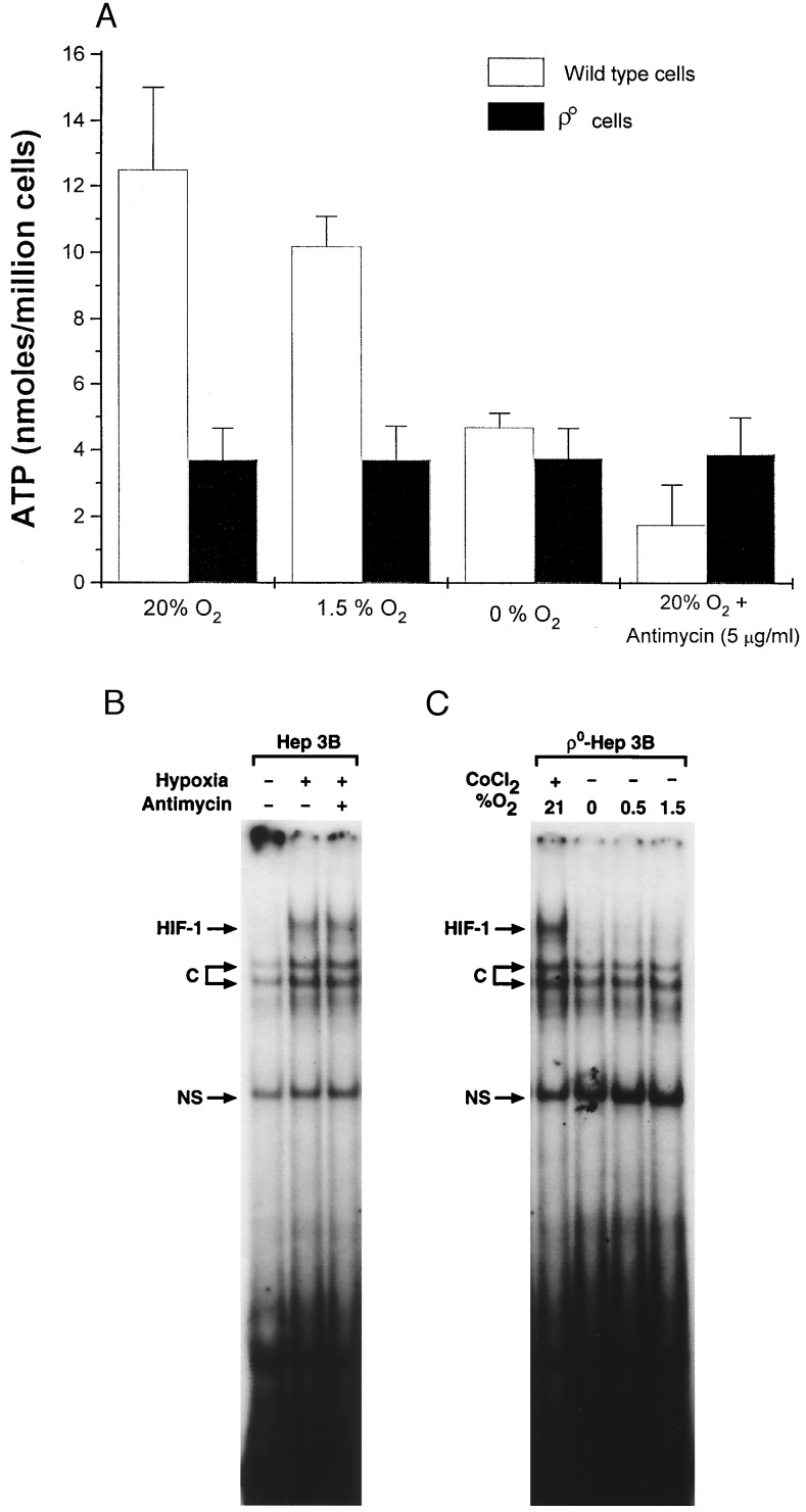
Figure 5.
(A) ATP levels (Boehringer Mannheim Bioluminescence Kit) in wild-type and ρ0-Hep3B cells during 21% O2, 1.5% O2, 0% O2, and antimycin (5 μg/ml) at 21% O2 for 1 hr. (B) HIF-1 DNA-binding activity in nuclear extracts from wild-type cells during hypoxia (1.5% O2/5% CO2/93.5% N2) in the presence of antimycin (5 μg/ml). (C) HIF-1 DNA binding in nuclear extracts from ρ0 cells during CoCl2 (100 μM), 1.5% O2, 0.5% O2, and 0% O2. C, constitutive; NS, nonspecific.

