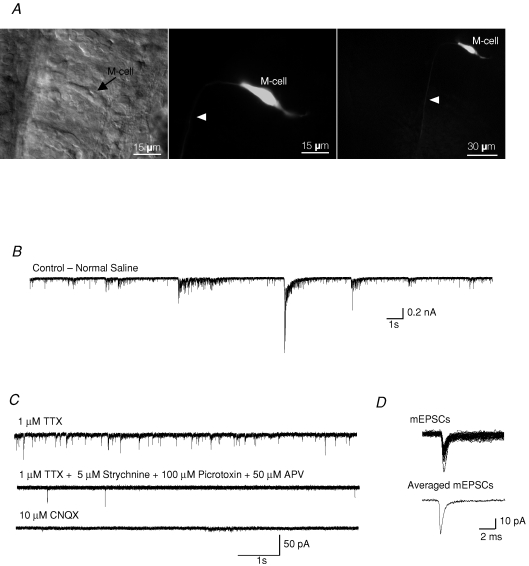
Figure 1.
Spontaneous synaptic activity in Mauthner cells (M-cells) A, differential interference contrast image of an M-cell in a hindbrain preparation. Arrow points to the cell body (left panel). The same M-cell was filled with Lucifer Yellow (0.1%) during a typical experiment (middle and right panel). Arrowhead points to the axon descending into the spinal cord. Scale bar: 15 μm in the left panel and the middle panel, and 30 μm in the right panel. B, spontaneous activity in an M-cell recorded in normal extracellular saline solution. C, miniature postsynaptic currents (mEPSCs) recorded in the presence of 1 μm TTX, 5 μm strychnine, 100 μm picrotoxin and 50 μm APV. All mEPSCs were completely blocked after bath application of the non-NMDA receptor antagonist CNQX (10 μm). D, individual mEPSCs were acquired (top) and averaged (bottom). Holding potential was −60 mV.

