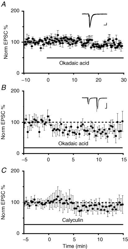
Figure 6.
Activation of phosphatase I and II are required for LTD A, average normalized EPSC amplitude is plotted against time for 6 cells in which the phosphatase I blocker okadaic acid was applied at time zero. Okadaic acid had no effect on basal transmission. The inset shows superimposed traces from one cell before and after application of okadaic acid. Scale bars are 100 pA and 10 ms B, average normalized EPSC amplitude is plotted against time in the presence of okadaic acid (1 μm) in the perfusing Ringer solution. The parabrachial input was stimulated at 1 Hz (900 pulses) at time 0 min (n = 8). LTD was absent in the presence of the protein phosphatase blocker. The inset shows superimposed traces from one cell before and after low-frequency stimulation. Scale bars, 50 pA and 10 ms C, similarly, incubation of slices in the protein phosphatase II blocker calyculin A (1 μm) also blocked the induction of LTD (n = 13).

