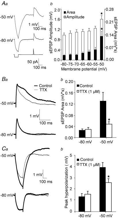
Figure 1.
Amplification of sEPSPs elicited by somatic current injection is abolished by bath-applied TTX Aa, EPSP-like depolarizations elicited by somatic current injection (lower traces, see methods) at −80 mV (middle traces), and at a depolarized potential near firing threshold (upper traces). The duration and area under the EPSP waveform were significantly increased by depolarization. The response to small hyperpolarizing steps was similarly enhanced. b, bar graph summarizing the effects of membrane potential on sEPSP amplitude and area. Single factor ANOVA (n = 10 cells) demonstrated significant effects of membrane potential on sEPSP amplitude (F6,63 = 4.92; P < 0.001) and sEPSP area (F6,63 = 7.44, P < 0.001). Amplitude and area at −50 mV were significantly different compared to those at −80 mV (*P < 0.05). Ba, example sweeps illustrating the attenuation of sEPSP amplification at depolarized potentials by TTX (1 μm). b, bar graph summarizing the effects of TTX (1 μm) on sEPSPs. Two factor ANOVA revealed significant effects of membrane potential (F1,36 = 14.65 P < 0.001), of TTX application (F1,36 = 5.93 *P < 0.02) and a significant interaction (F1,36 = 7.09, P < 0.02). Ca, example sweeps illustrating the effects of TTX (1 μm) on the response to hyperpolarizing steps elicited at hyperpolarized and depolarized potentials. b, bar graphs summarizing the effects of TTX (1 μm) on the response to hyperpolarizing steps (n = 10 cells). Two factor ANOVA revealed significant effects of membrane potential (F1,36 = 32.83 P < 0.001), and a significant interaction (F1,36 = 6.15 *P = 0.02), suggesting that the effects of TTX were conditional to membrane potential value.

