
Fig. 3.
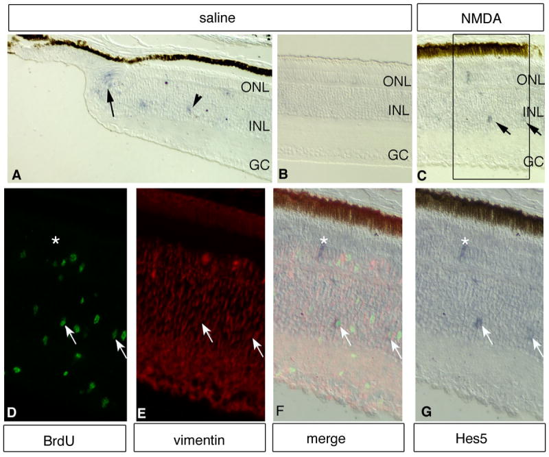
Hes5 is expressed in the de-differentiating, proliferating Müller glia. Intraocular injections of saline (A, B) or NMDA (C-G) were made at P1, BrdU was injected at P3 and birds were sacrificed at P4. Sections were treated by in situ hybridization with a probe against Hes5. (A) Peripheral region of control (saline injected) retina. Arrows point to Hes5 expressing cells in the CMZ. (B) Central region of control (saline injected). Hes5 expression is not detected in the central retina of saline injected eyes. (C) Central region of NMDA injected retina. (D-G) Enlargement of boxed area shown in C. Several de-differentiating, proliferating Müller glia express Hes5. Arrows point to cells stained positive for BrdU (green), Notch, and vimentin (red). Asterisk marks a cell stained positive for Hes5 but not BrdU.