
Fig. 7.
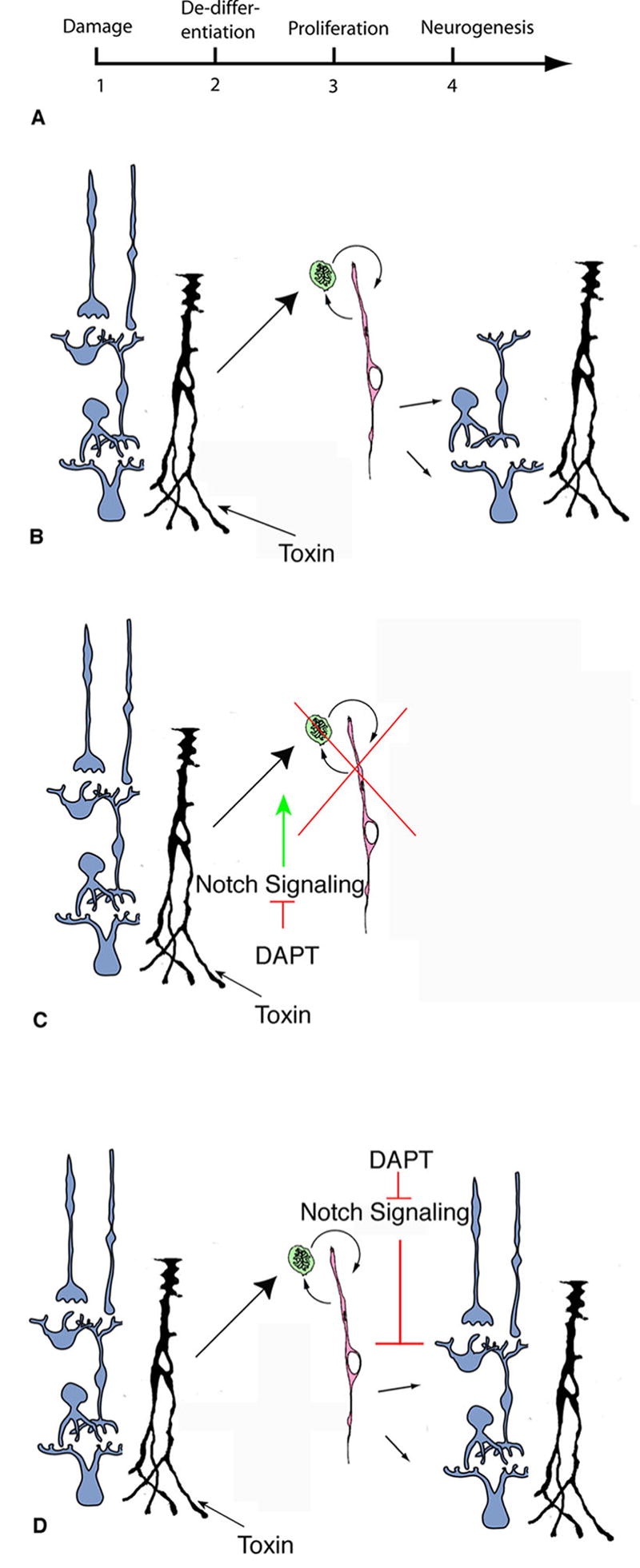
DAPT can affect the regenerative process at two distinct phases. (A) Timecourse of regeneration. (B) Schematic of regeneration. A toxin, such as NMDA, causes Müller glia (black cells) to respond by proliferating (green cells) and de-differentiating in progenitor cells (pink cell). A subset of the progenitor cells differentiates into new neurons. (C) If DAPT is injected early, as Müller cells are dedifferentiating into progenitor cells, there is less dedifferentiation and proliferation, indicating that Notch signaling promotes this transition. (D) If DAPT is injected later, after Müller cells have dedifferentiated into progenitor cells, there are more newly generated neurons, indicating that Notch signaling may normally prohibits this transition.