
Fig. 1.
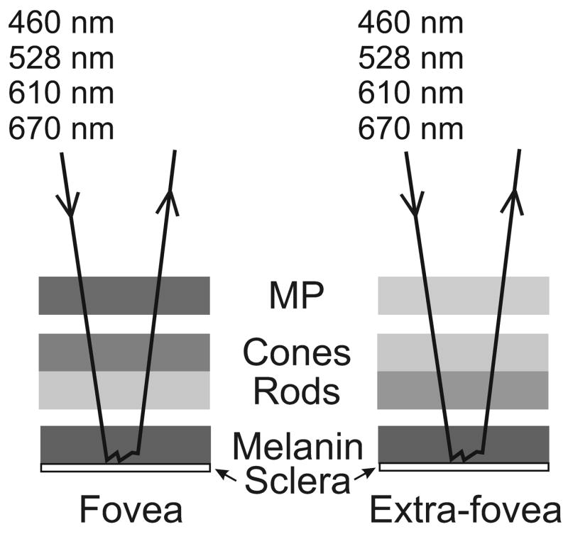
Model of the retina as a sequence of absorbing layers: macular pigment (MP), cone and rod photopigments, and melanin. Incident light, of each of four wavelengths, is absorbed to a greater or lesser extent by each layer and the amount each layer absorbs is generally different depending on retinal location. The light is returned through the layers after multiple scattering in the melanin layer and/or reflection at the sclera.