
Fig. 1.
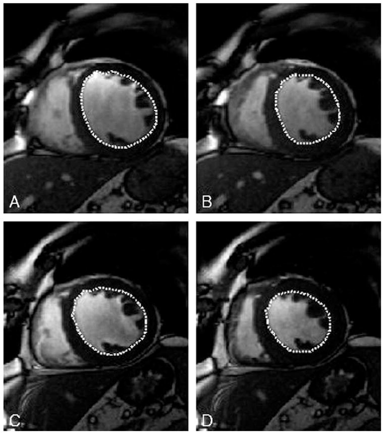
Assessment of left-ventricular volumes by cine-MRI. Endo- and epicardial borders were manually traced in all slices in end-diastole and end-systole, papillary muscles were traced separately. After administration of oxypurinol, MR imaging was repeated with corresponding slice orientations. (A) Diastolic endocardial borders in a midventricular slice before administration of oxypurinol. (B) Systolic endocardial borders in a midventricular slice before administration of oxypurinol. (C) Diastolic endocardial borders in a midventricular slice after administration of oxypurinol. (D) Systolic endocardial borders in a midventricular slice after administration of oxypurinol.