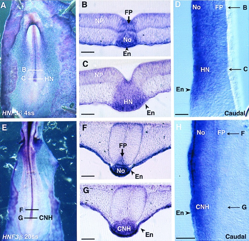
Figure 2.
HNF3β expression pattern in 4ss (A–D) and in 20ss (E–H) chicken embryos. (A and E) In toto ventral views. (B, C, F, and G) Transverse sections. HNF3β transcripts are present in HN-CNH and in the floor plate (FP) and notochord (No) immediately rostral to HN-CNH. The gene is slightly more strongly expressed in the vicinity of the endoderm (En), where it is also expressed. Sagittal sections show the continuity between Hensen’s node (HN) and FP/No at 4ss (D) and between CNH and FP/No at 20ss (H). NP, neural plate. (Bars = 80 μm.)

