
Fig. 13.
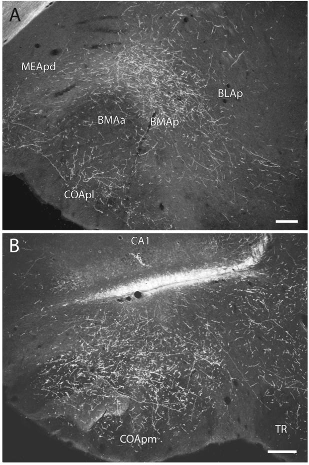
Darkfield photomicrographs showing the distribution of PHAL-labeled axons within the posterior basomedial amygdalar nucleus (A) and the posteromedial cortical amygdalar nucleus (B) following an injection centered in ventral field CA1 (Experiment HIPPO161). Scale = 200 μm.