
Figure 2.
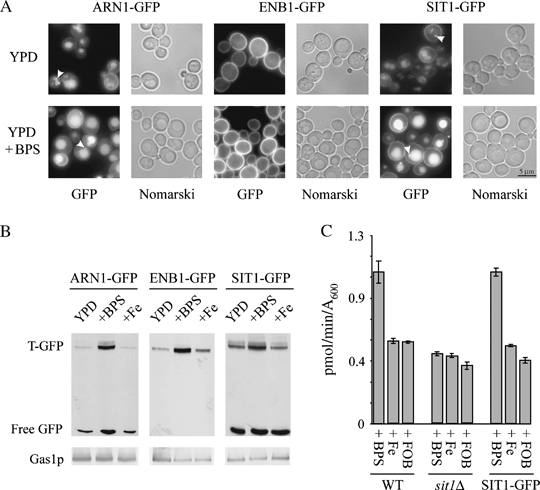
Synthesis and location of chromosomal GFP-tagged siderophore transporters.A) Cells expressing chromosomal GFP-tagged siderophore transporters were cultured to midexponential growth phase in complete medium (YPD) or in complete medium supplemented with 200 μm BPS for 4 h (YPD + BPS). Cells were then examined for GFP fluorescence and with Nomarski optics. Arrows indicate round structures possibly corresponding to endosomes. B) Total protein extracts were prepared and analysed by Western blotting for GFP [GFP-tagged transporter (T-GFP) and free vacuolar GFP] and for Gas1p, as a loading control. C) We determined FOB uptake by WT, sit1Δ and SIT1-GFP cells cultured to midexponential growth phase in complete medium supplemented with 200 μm BPS, 100 μm iron citrate (Fe) or 100 μm FOB (means ± standard error of the mean from three experiments). WT, wild type.