Figure 9.
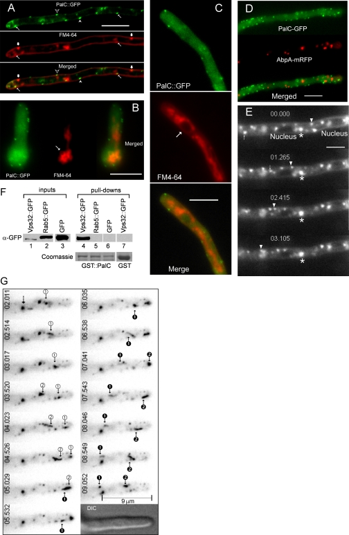
Characterization of PalC-GFP and Vps32-GFP-containing compartments.A) PalC-GFP punctate structures and FM4-64-stained plasma membrane and associated cortical structures stained shortly after dye loading. Examples of cortical and nearly cortical PalC-GFP punctate structures that do not associate with cortical FM4-64 puncta are indicated by empty and filled arrowheads, respectively. Thick arrows indicate two examples of FM4-64 cortical structures that do not associate with PalC-GFP. Thin arrows indicate two (rare) examples where PalC-GFP and FM4-64 cortical structures are closely associated. B and C) PalC-GFP punctate structures do not associate with endomembranes stained with FM4-64. B) One of several vacuoles labelled by FM4-64 after a 45-minute chase (see text) in the swelled basal conidiospore is arrowed. C) Endomembranes stained with FM4-64 after a 45-minute chase include a network of mitochondria and endoplasmic reticulum (ER). One nucleus showing labelling of its ER-associated membrane is arrowed. D) PalC-GFP punctate structures do not associate with AbpA-mRFP-labelled actin patches. E) Motile FM4-64 early endosomes filmed within 5 minutes after dye loading in an incubation chamber at 28°C. Frames were taken from Movie S2, which was made using the stream acquisition feature of the MetaMorph Universal Imaging software with 0.1 seconds exposures. Elapsed time is indicated in seconds:milliseconds. One of the several endosomes moving very near the cortex is indicated with an arrowhead. A second endosome moving in the opposite direction and stopping in the vicinity of a nucleus is arrowed. A larger, static endosome is indicated with an asterisk. Bar, 5 μm. F) GST-PalC pulls down Vps32-GFP but not GFP-Rab5 or GFP from Aspergillus nidulansprotein extracts from cells expressing the indicated proteins. Glutathione S-transferase does not pull down Vps32-GFP. Glutathione–Sepharose bound proteins were run in twin 10% polyacrylamide gels, one of which was analysed by Western blot using an anti-GFP antibody (α-GFP), whereas the second was stained with Coomassie Blue to show the amounts of GST-PalC and GST baits used in the different lanes. G) Motile endosomes labelled with Vps32-GFP. Frames, shown in inverted contrast, were taken from Movie S3, which should be consulted to get a more accurate record of endosome motility and trajectories. The movie was made using the stream acquisition feature of MetaMorph with 0.5 seconds exposures and a 2 × 2 binning. Elapsed time is indicated in seconds:milliseconds. Endosomes showing retrograde and anterograde movement (two of each class) are indicated with arrowed numbers. Note that subapical ‘black’ endosome 2 appears to be formed after receiving traffic from ‘white’ endosomes 1 and 2 moving in anterograde direction. The dotted arrow indicates a static endosome, which may be used as reference.

