Figure 5.
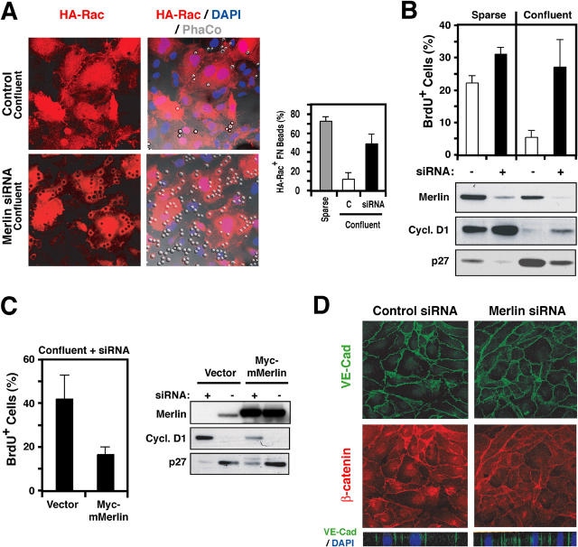
Merlin mediates contact inhibition of growth by suppressing membrane recruitment of Rac. (A) HUVEC were electroporated with GFP-Rac and then either transfected with a siRNA oligonucleotide targeting human Merlin or mock-transfected as a control. 36 h later, cells were synchronized in G0, detached, and plated on FN under either sparse or confluent conditions, as indicated, and treated with FN beads. The graph shows the percentage of GFP-Rac–positive beads under the indicated conditions. (B) Cells were transfected with a siRNA oligonucleotide targeting human Merlin (+). G0-synchronized cells were detached, plated on FN under sparse or confluent conditions, and incubated with mitogens and BrdU for 20 h. The graph shows the percentage of cells entering S phase under the indicated conditions. Total lysates were subjected to immunoblotting with the indicated antibodies. (C) Cells were electroporated with a vector encoding Myc-tagged mouse Merlin (mMerlin) or empty vector and transfected with the anti–human siRNA oligonucleotide. After 36 h, cells were synchronized in G0, detached, and plated on FN under confluent conditions for 4 h. The cells were either incubated with mitogens and BrdU for 20 h to measure entry into S phase or treated with mitogens for 12 h and subjected to immunoblotting with the indicated antibodies. (D) siRNA-transfected cells were detached and plated on FN-coated coverslips under confluent conditions in the presence of growth factors. After 20 h, they were subjected to double immunofluorescent staining with anti–VE-cadherin (green) and anti–β-catenin (red). The bottom panels are XZ section views of VE-cadherin and DAPI staining. Error bars represent the mean ± SD.

