
Figure 9.
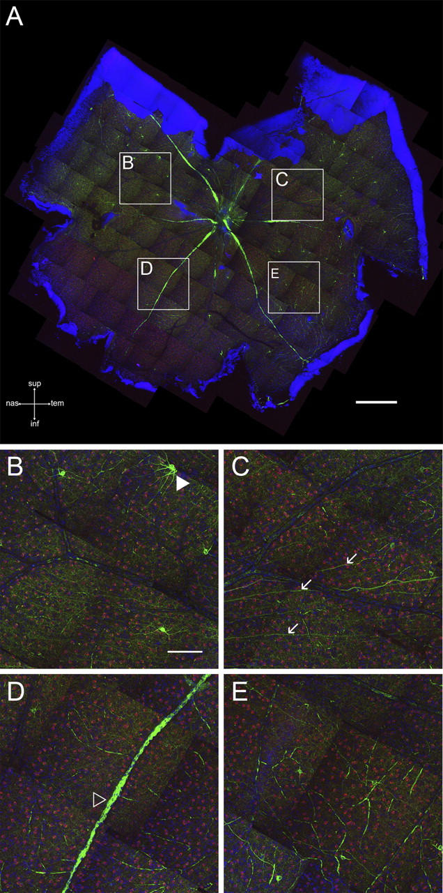
Whole mount of a severely affected retina with virtually no remaining ganglion cells. (A) High-resolution survey of a whole-mounted retina from one of the most severely affected eyes in our sample stained for SMI32 (green), ChAT (red), and counterstained with TOPRO (blue). Bar, 500 μm. (B–E) High-power views of the boxed areas outlined in A. Hardly any SMI32+ cells or axons are left. Remaining cells often show long, unbranched dendrites. The open arrowhead in D points to a blood vessel that has been unspecifically labeled by the secondary antibody. The closed arrowhead in B points to an anomalous SMI32+ cell. The arrows in C indicate remaining axons. Note that the ChAT+ cells (red) are unaffected. Bar, 100 μm.