Figure 1.
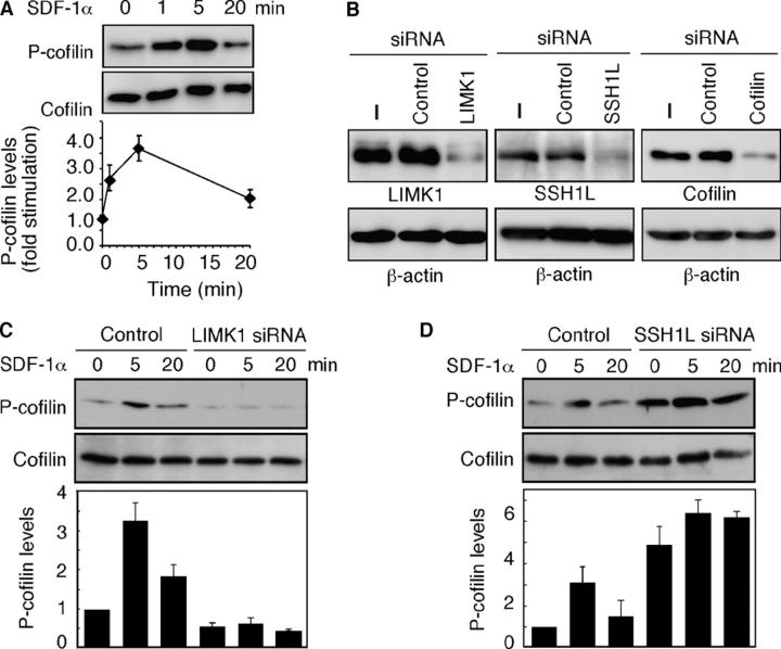
SDF-1α–induced changes in P-cofilin levels are regulated by LIMK1 and SSH1L. (A) SDF-1α–induced changes in P-cofilin levels. Jurkat cells were stimulated with 5 nM SDF-1α for the indicated times, and cell lysates were analyzed by immunoblotting with anti–P-cofilin and anticofilin antibodies. The bottom panel shows the relative P-cofilin levels after SDF-1α stimulation as means ± SEM of triplicate experiments. (B) Suppression of endogenous LIMK1, SSH1L, and cofilin expression by siRNA. Jurkat cells were transfected with siRNA plasmids for GFP (control), LIMK1, SSH1L, cofilin, or empty vector (−). After 60 h of culture, cell lysates were analyzed by immunoblotting with antibodies specific for each protein and β-actin. For LIMK1 and SSH1L, the cell lysates were subjected to immunoblotting after immunoprecipitation. (C and D) Effects of LIMK1 or SSH1L siRNA on SDF-1α–induced changes in P-cofilin levels. SSH1L, LIMK1, or GFP (control) siRNA cells were stimulated with 5 nM SDF-1α. Cell lysates, prepared at the indicated times, were analyzed by immunoblotting as in A. The bottom panels indicate the relative P-cofilin levels; the value at time = 0 in control cells is taken as 1.0. Each value represents the mean ± SEM (error bars) of triplicate experiments.

