
Figure 3.
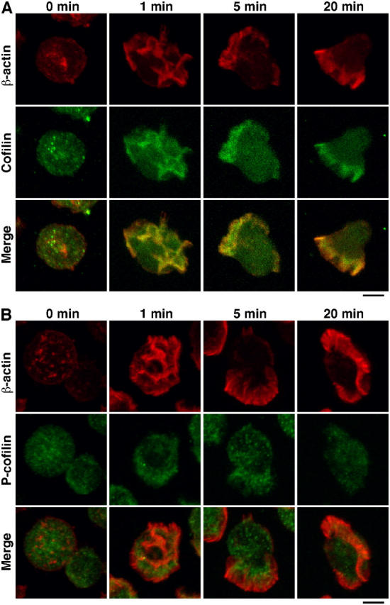
Cofilin, but not P-cofilin, accumulates in the SDF-1α–induced lamellipodial membrane protrusion in Jurkat cells. Jurkat cells were left unstimulated (0 min) or were stimulated with 5 nM SDF-1α for the indicated periods of time. Cells were costained with anti–β-actin mAb (red) and anticofilin (A) or anti–P-cofilin (B) pAbs (green). Merged images are shown in the bottom panels. Bars, 5 μm.