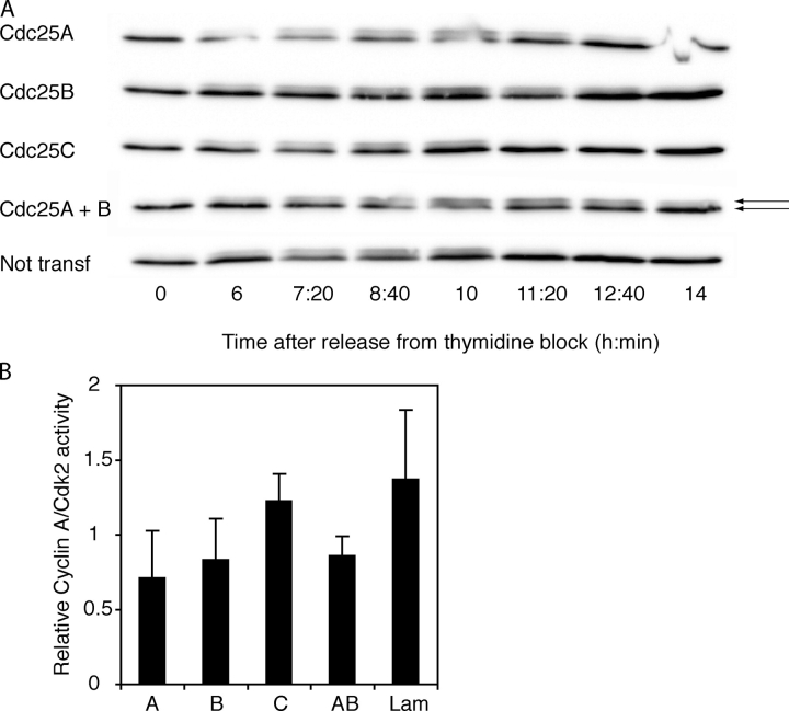
Figure 6.
Reduced activities of both cyclin A–Cdk2 and cyclin B1–Cdk1 in lysates of cells transfected with siRNA to Cdc25A or -B. (A) Delayed dephosphorylation of Cdk1 in cells treated with siRNA to Cdc25A or -B. SiRNA-transfected synchronized cells were subjected to Cdk1 immunoblotting. Arrows indicate the faster migrating unphosphorylated Cdk1 (bottom band) and the slower migrating phosphorylated Cdk1 (top band). siRNAs are indicated to the left. A quantification of the ratios of inactive versus active Cdk1 is available in Fig. S1 (available at http://www.jcb.org/cgi/content/full/jcb.200503066/DC1). (B) Reduced activation of cyclin A–Cdk2 in cells treated with siRNA to Cdc25A or -B. Cyclin A was immunoprecipitated from siRNA-transfected cells 9 h after release from thymidine block. The ability of the immunoprecipitate to phosphorylate histone H1 as well as the amount of Cdk2 in the immunoprecipitate was assessed. Bars show average from three independent experiments of normalized ratio between cyclin A–Cdk2 activity and amount of Cdk2.

