
Figure 5.
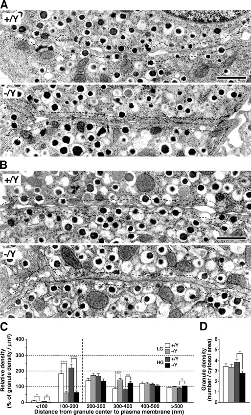
Granule docking in isolated islets. (A and B) Electron micrographs of β cell sections. Islets were isolated from 24-wk-old male Grn + /Y and Grn − /Y mice and incubated at 37°C with 2.8 mM LG buffer for 1 h (A) and then with 25 mM HG buffer for 30 min (B). Dashed lines indicate a border 200 nm distant from the plasma membrane. Bars, 1 μm. (C and D) Morphometric analyses of insulin granules in LG-treated Grn + /Y (white bars) and Grn − /Y islets (light gray bars), and HG-treated Grn + /Y (dark gray bars) and Grn − /Y islets (black bars). For each group, 10 randomly selected β cells from four different animals were analyzed. (C) Relative density of insulin granules located near the plasma membrane. The granules were categorized into six bins according to their distance from the granule center to the plasma membrane (nm). The data were represented as a percentage of the granule density in each bin (100% corresponds to the average granule density in cytoplasm). *, P < 0.05; **, P < 0.005; ***, P < 0.0003. (D) Average granule number per cytosol area (μm2). *, P = 0.012. Results are provided as mean ± SEM.