
Figure 1.
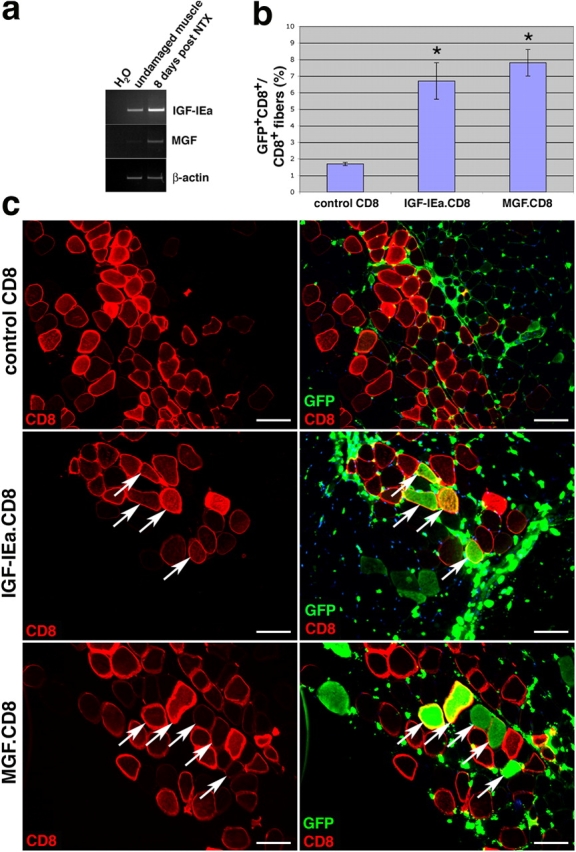
DNA electrotransfer of IGF-I increases the contribution of BMDCs to skeletal muscle. (a) The two muscle-specific isoforms, IGF-IEa and MGF, are shown to be up-regulated in the muscle after notexin damage as shown by RT-PCR analysis. (b) Plasmids encoding IGF-IEa.CD8, MGF.CD8, or CD8 only (control) were electroporated into the TA muscles of mice that had received a BMT 8 wk earlier, and muscles were harvested 4 wk after DNA delivery. The percentage of GFP+CD8+ myofibers relative to total CD8+ myofibers is shown (± SEM). P value was determined with a t test. *, P < 0.02. (c) Immunofluorescence images of transverse sections of muscle stained for CD8 only (left) or for GFP and CD8 (right). GFP+ cells <10 μm are blood cells. Arrows indicate examples of CD8+ myofibers that were also GFP+. Bars, 50 μm.