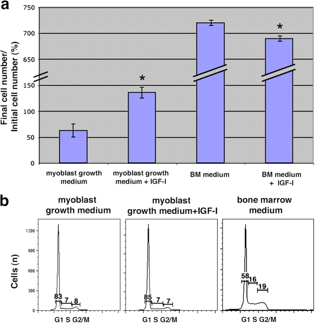
Figure 6.
IGF-I promotes survival of MMCs. (a) MMCs were obtained by FACS from freshly isolated bone marrow and 6 × 10⁴ cells plated in myoblast GM for 3 d, with or without 100 ng/ml IGF-I. Cell counts were performed on days 0 and 3. Final cell numbers are expressed as a percentage of the initial number of cells plated on day 0 (± SEM). P value was determined with a t test. *, P < 0.02. (b) Cell cycle profile using propidium iodide for analysis of DNA content in MMCs with or without IGF-I after 3 d of culture in the media indicated. Percentages of the cells in each specific cell cycle phase are indicated on the graphs.

