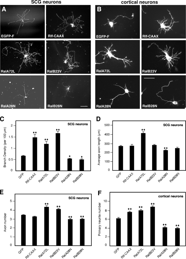
Figure 1.
Active Ral increases neurite branching. SCG neurons (A) and cortical neurons (B) expressing EGFP-F or the indicated proteins visualized by anti-myc (for Ral) or anti-HA (for Rlf-CAAX) immunostaining. Pictures were taken 20 h after transfection. Constitutively active RalA (RalA72L) and -B (RalB23V) or the active Ral-GEF Rlf-CAAX increase neurite branching, whereas dominant-negative Ral (RalA28N and -B28N) decreases branching. SCG neurons were plated on laminin-coated dishes, whereas cortical neurons were grown on polyornithine. Bars: (A) 100 μm; (B) 50 μm. (C and D) Quantitative analysis of neurite morphology. Branch density per 100 μm of neurite length (C) is highly increased with Rlf-CAAX and constitutively active Ral and decreased with dominant-negative Ral (means ± SEM: GFP, 0.65 ± 0.03; Rlf-CAAX, 1.45 ± 0.14; RalA72L, 1.17 ± 0.12; RalB23V, 1.62 ± 0.09; RalA28N, 0.52 ± 0.03; RalB28N, 0.48 ± 0.03; **, P < 0.0001; *, P < 0.01). In contrast, average axon length (D) does not vary significantly, except for Ral A (means ± SEM: GFP, 261.62 ± 8.65; Rlf-CAAX, 266.41 ± 16.92; RalA72L, 402.54 ± 18.57; RalB23V, 274.53 ± 9.01; RalA28N, 216.89 ± 7.92; RalB28N, 238.74 ± 14.52; **, P < 0.0001). (E and F) Quantitative analysis of the number of primary neurites emerging from the cell body and containing microtubules in SCG neurons (means ± SEM: GFP, 3.38 ± 0.06; Rlf-CAAX, 3.20 ± 0.07; RalA72L, 4.25 ± 0.17; RalB23V, 4.03 ± 0.11; RalA28N, 2.91 ± 0.10; RalB28N, 2.93 ± 0.10; **, P < 0.0001) and cortical neurons (means ± SEM: GFP, 5.95 ± 0.23; Rlf-CAAX, 7.33 ± 0.18; RalA72L, 7.67 ± 0.26; RalB23V, 8.42 ± 0.25; RalA28N, 3.98 ± 0.20; RalB28N, 3.78 ± 0.18; **, P < 0.0001).

