Figure 4.
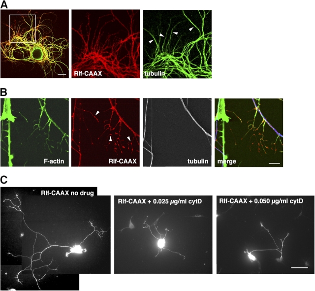
Branches induced by Ral activation depend on F-actin. (A, left) Confocal image of an SCG neuron plated on polyornithine expressing Rlf-CAAX (red) and stained for tubulin (green). The middle and right panels are enlargements of the boxed area in the left panel. Longer branches are positive for tubulin (arrowheads). Bar, 20 μm. (B) In Rlf-CAAX–expressing neurons, short branches contain actin filaments visualized by fluorescent phalloidin (green) but not tubulin (white, blue in the merge panel). Note the accumulation of Rlf-CAAX (red) in discrete domains along branches (arrowheads). Bar, 10 μm. (C) Representative images of SCG neurons plated on polyornithine, expressing Rlf-CAAX, and treated with the indicated amounts of cytochalasin D. Increasing drug doses led to a progressive block of Ral-dependent branching. Bar, 50 μm.

