
Figure 4.
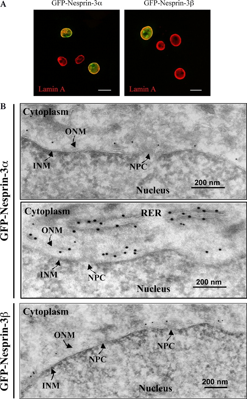
Subcellular localization of nesprin-3α and -3β. (A) To determine the subcellular location of nesprin-3, PA-JEB keratinocytes were transiently transfected with either GFP-nesprin-3α or -3β cDNA, and the expression pattern was analyzed by fluorescence microscopy. Immunofluorescence studies of endogenous lamin A were performed using mAb 133A2. Bar, 20 μM. Four cells are shown in each image. (B) To determine which lipid bilayer of the NE contains nesprin-3, ultrathin sections of PA-JEB cells stably expressing either nesprin-3α (top and middle) or -3β (bottom) were labeled with rabbit pAbs against GFP, followed by incubation with 15-nm gold-conjugated protein A. NPC, nuclear pore complex.