
Figure 2.
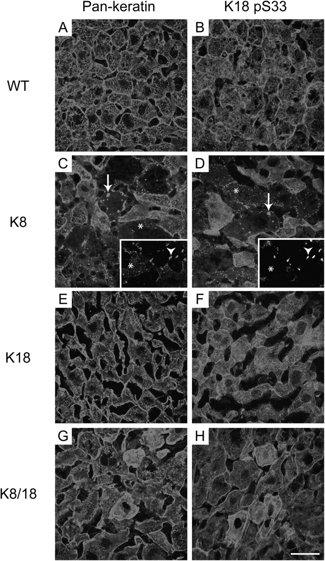
Spontaneous pre-MB formation in livers of old K8 mice. Immunofluorescence staining was performed on livers of 2-yr-old WT, K8, K18, and K8/18 mice. Livers were stained with Abs to K8/18 (pankeratin; A, C, E, and G) or to K18 pS33 (B, D, F, and H). Arrows (C and D) highlight keratin aggregates that are seen only in K8-overexpressing mice, whereas the asterisks highlight fine keratin dots that are likely precursors to the larger aggregates. Bar, 50 μm. Insets show double staining of K8/18 (C) and ubiquitin (D) and illustrate that the larger keratin aggregates are ubiquitin positive (arrowheads), whereas the smaller keratin dots are ubiquitin negative (asterisks).