
Figure 5.
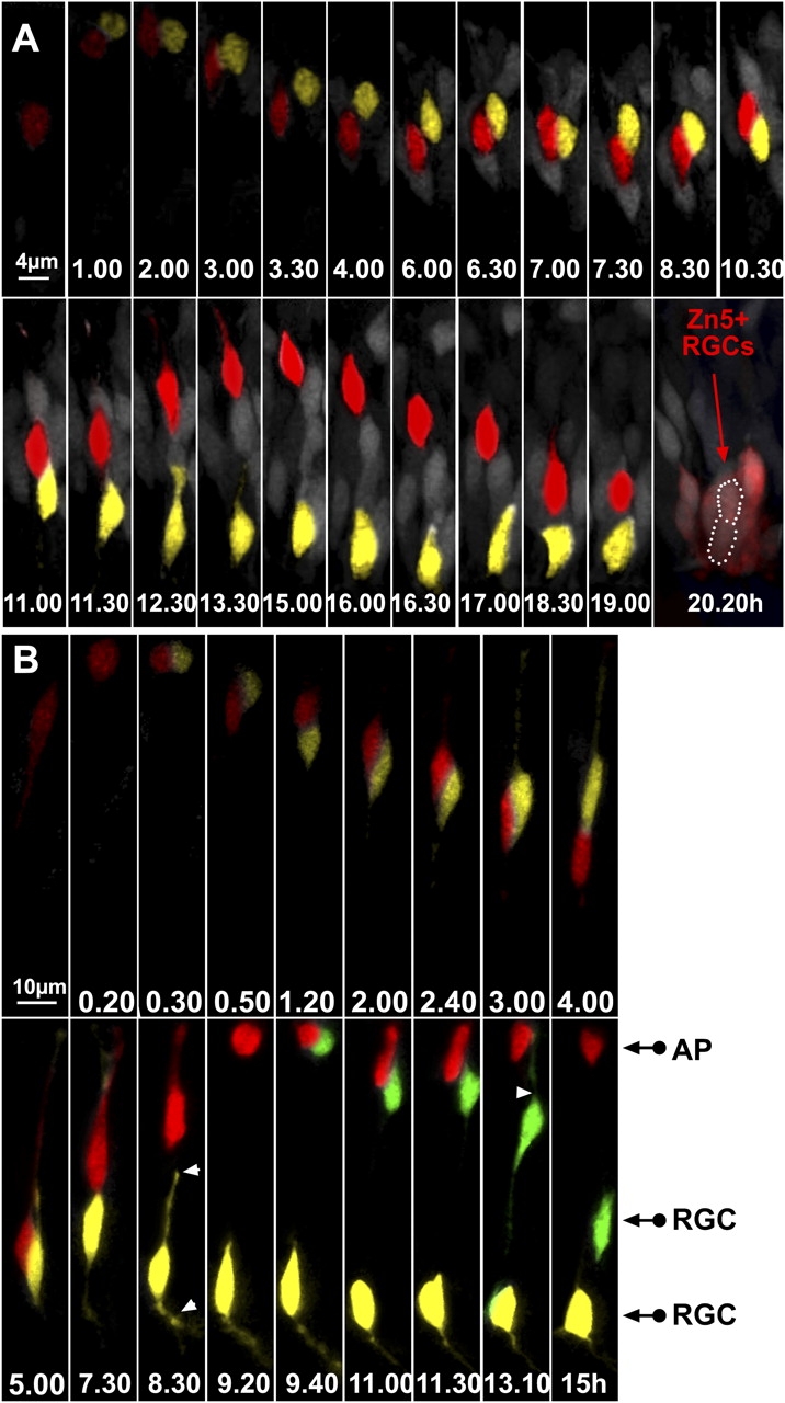
ath5:GFP progenitors generate two RGCs after division in the lakritz environment. (A) The two daughter cells have been highlighted in yellow or red. Time-lapse series showing an example of an ath5:GFP progenitor generating two RGCs in the lakritz environment. Imaging was started at 30–32 h after fertilization, and t = 0 corresponds to the time of appearance of ath5:GFP (4 h after the onset of the video recording). After 11 h, the red daughter cell starts migrating toward the apical surface. Once it has reached the apical surface (t = 15 h), it migrates back again toward the basal surface, where it differentiates in RGCs. The location of both daughter cells after 20 h is outlined by a white dotted line. Both cells were zn-5 positive after immunolabeling of the imaged retina. (B) An example of a time-lapse series showing an ath5:GFP progenitor that divides and generates another dividing progenitor. Imaging was started 30–32 h after fertilization, and t = 0 corresponds to the time of appearance of ath5:GFP (3 h after the onset of the video recording). At t = 9.40 h, the progenitor highlighted in red divides once more at the apical surface, generating one daughter cell (green) that lost its apical process and began to put out an axon and another daughter cell (red) that remained apical. White arrowheads point to the retracting apical process and the forming axon. AP, apical cell; RGC, retinal ganglion cell.