
Figure 1.
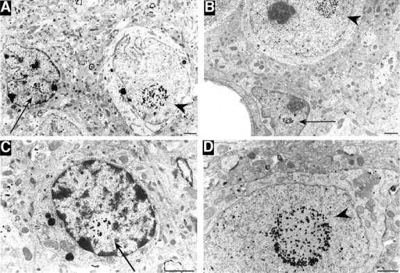
Electron micrographs of HD mouse brains. (A–D) EM48 immunogold labeling of the striatum (A) and cortex (B–D) of R6/2 mice at 12 wk of age. Immunogold-labeled aggregates are present in glial cells (arrows), which show a more condensed nuclear membrane and a smaller sized cytoplasm than do neuronal cells (arrowheads). Bars, 1 μm.