Figure 2.
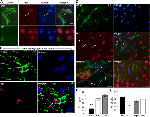
Immunofluorescent labeling of htt-containing glia in R6/2 transgenic mouse brains. (A) Immunofluorescent labeling of brain sections containing white matter (WM) and the central region of the cerebellar cortex (Ctx) in an R6/2 mouse 8 wk old. Mouse antibody to GFAP labeled astrocytes (green) and rabbit EM48 labeled mutant htt (red). The nuclei were labeled with Hoechst dye (blue). (B) Confocal imaging of white matter showing that the nuclei of GFAP-positive glial cells contain htt aggregates (Video 1, available at http://www.jcb.org/cgi/content/full/jcb.200508072/DC1). (C) The striatal section of an R6/2 mouse at 8 wk of age was stained with antibody to GFAP and the nuclear dye Hoechst (top). EM48 immunostaining for htt and merged image are also presented (middle). Bottom panels shows merged images of striatal regions from 12- or 4-wk old R6/2 mice. Arrows indicate glial nuclei that contain EM48 staining. Small puncta represent neuropil aggregates. (D) The percentage of glial cells containing nuclear EM48 staining in the brain striatal sections of R6/2 mice at the age of 4, 8, or 12 wk. (E) The percentage of glial cells with intranuclear htt in the striatum (Str), cortex (Ctx), hippocampus (Hipp), and WM in R6/2 mice at the age of 8–10 wk. The data (mean ± SD) were obtained from three to five mice per group. **, P < 0.01 compared with the samples of 8 or 12 wk old R6/2 mice. Bars: (A) 5 μm; (B) 2.5 μm; (C)10 μm.

