Figure 4.
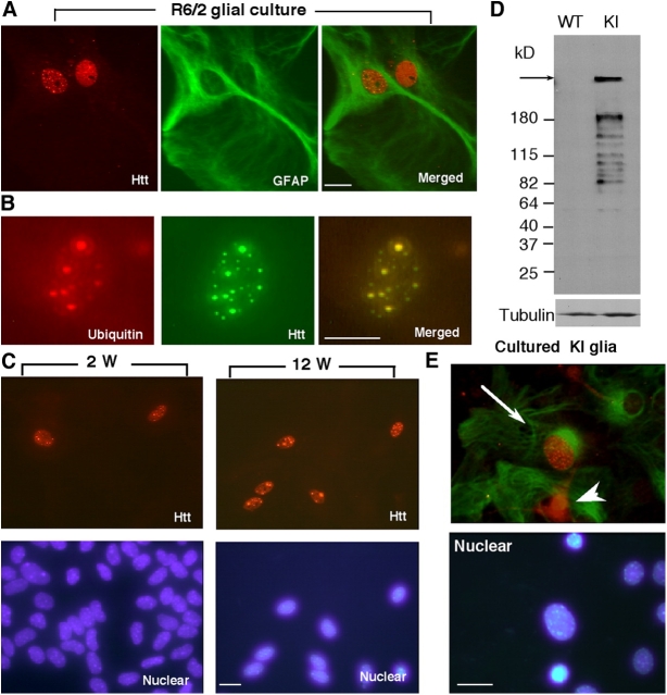
Age-dependent nuclear accumulation of mutant htt in cultured glial cells. (A) Immunofluorescence double labeling showing that GFAP (green) positive astrocytes display htt aggregates (red) in the nuclei of R6/2 glial cells. (B) Some intranuclear htt aggregates are also labeled by antibody to ubiquitin. (C) Immunofluorescent images of cultured glial cells that were cultured for 2 and 12 wk showing the increase of glial htt aggregates with time. (D) Western blotting of cultured astrocytes that were isolated from the cortex of postnatal Hdh CAG(150) knock-in (KI) and littermate control (WT) mice and had been cultured for 4 wk. The blot was probed with antitubulin (bottom) and 1C2 (top), an antibody that is specific to expanded polyQ tracts and reacts with NH2-terminal htt fragments containing 150Q. Arrow indicates full-length mutant htt. (E) Immunofluorescent images of glial culture from Hdh CAG KI. GFAP (green) positive astrocytes (arrows) contain intranuclear htt (red) aggregates. Some GFAP-negative cells (arrowhead) also show intranuclear htt, suggesting that they might be immature astrocytes or other types of cells. Bars, 5 μm.

