
Figure 8.
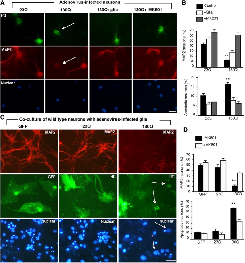
Htt-mediated neurotoxicity in glia–neuron coculture. (A) Cultured cortical neurons were infected with adenoviral htt-23Q or htt-130Q for 24 h. The infected neurons were then cultured with or without wild-type glial cells or MK801 (10 μM) and labeled by antibodies to htt (top), MAP2 (middle), and Hoechst (bottom). Htt-130Q–infected neurons show decreased MAP2-staining (arrows). (B) The percentage of MAP2-positive neurons and apoptotic neurons with nuclear DNA fragmentation in the presence or absence of wild-type glial cells or MK801. (C) Cultured glial cells (4–6 wk) infected with adenoviral htt-23Q or htt-130Q were cocultured with wild-type cortical neurons. EM48 immunofluorescence staining of glia–neuron coculture shows that htt-23Q is distributed in the cytoplasm whereas htt-130Q accumulates in the nuclei (arrows) of infected glial cells. The size of nuclei of cultured glial cells is often larger than that of cultured cortical neurons. There is a decrease in the number of MAP2-positive neurons in the coculture with htt-130Q–infected glial cells. Nuclei were stained with Hoechst (blue). (D) The percentage of MAP2-positive neurons and apoptotic cells in the presence of adenoviral infected glial cells. Neurons were treated with or without MK801 (10 μM). The data (mean ± SEM) were obtained by counting the number of degenerated cells and the total number of nuclei per image. **, P < 0.01 compared with neurons cocultured with glial cells. Bars, 10 μm.