
Figure 3.
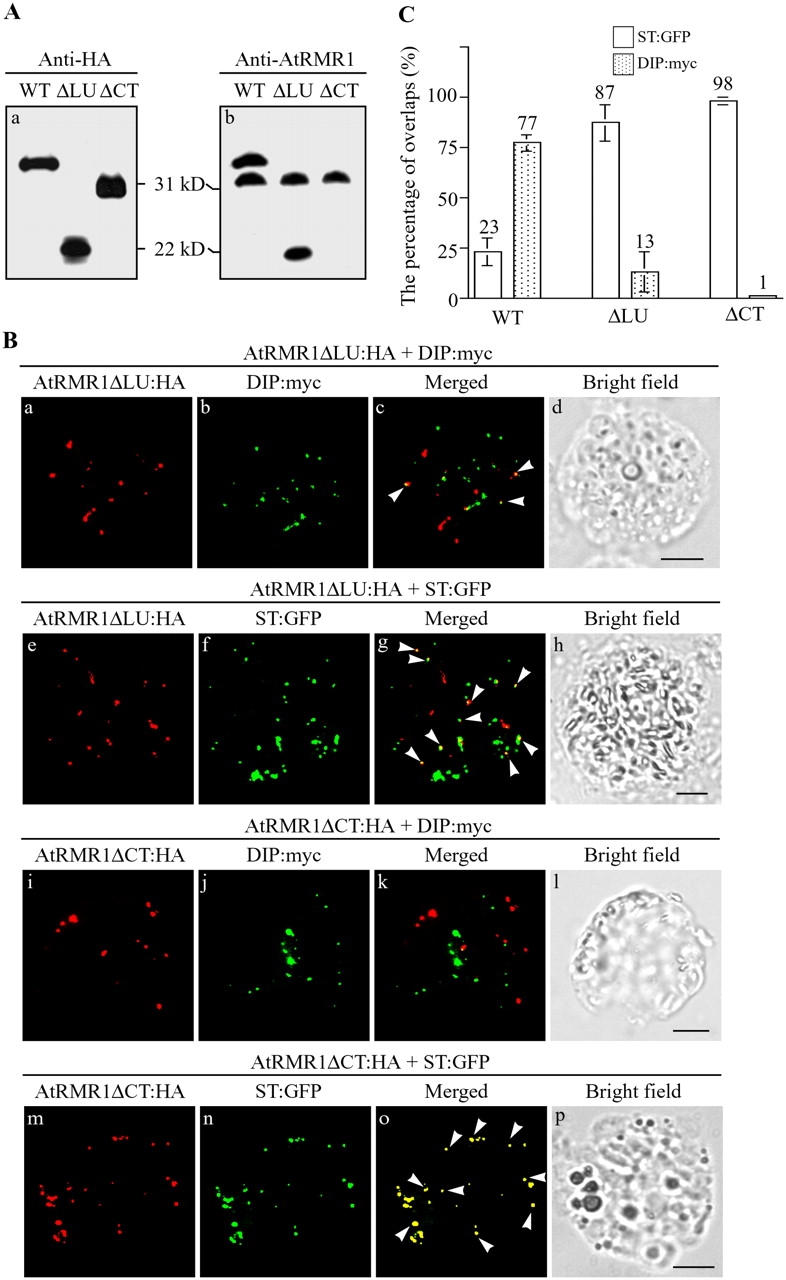
Localization of AtRMR1 deletion mutants. (A) Expression of AtRMR1 deletion mutants. Protein extracts were obtained from protoplasts transformed with the indicated constructs and subjected to Western blot analysis using anti-HA and anti-AtRMR1 antibodies. Note that anti-AtRMR1 antibody does not recognize AtRMR1ΔCT. (B) Localization of AtRMR1 deletion mutants. Protoplasts transformed with the indicated constructs were fixed and stained with anti-HA or anti-myc antibodies. The green fluorescent signal of ST-GFP was directly observed from fixed protoplasts. Arrowheads indicate overlaps between the indicated proteins. Bars, 20 μm. (C) Quantification of the overlaps between AtRMR1 deletion mutants and ST-GFP or DIP-myc. The number of punctate stains of AtRMR1 deletion mutants and ST-GFP or DIP-myc that overlapped were counted to determine localization of AtRMR1 deletion mutants. More than 200 punctate stains of AtRMR1 deletion mutants were counted for each comparison in a triplicate experiment. Error bars represent SEM. WT, wild-type AtRMR1; ΔLU, AtRMR1ΔLU-HA; ΔCT, AtRMR1ΔCT-HA.