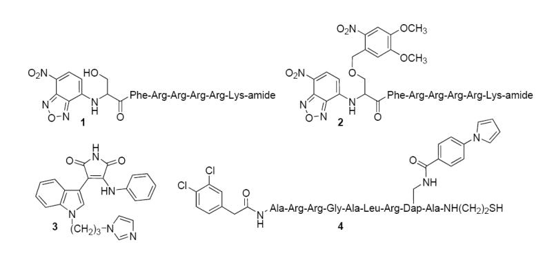
Figure 1.

Structures of PKC sensors and inhibitors. Compound 1 responds to PKC-catalyzed phosphorylation in a fluorescently sensitive fashion (7). The nonphosphorylatable analogue 2 is converted to the active sensor 1 by photolysis (8). Compound 3 is a selective PKC ß inhibitor (11), whereas compound 4 serves as a selective PKC α inhibitor (12).
