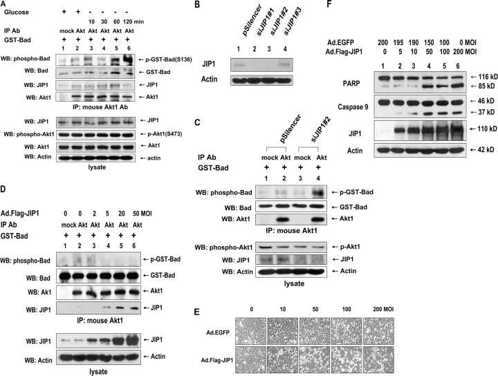
Figure 1.
Role of JIP1 in Akt activity in DU-145 cells. (A) Cells were exposed to glucose-free medium for various times (10–120 min). Cells were lysed, and lysates were immunoprecipitated (IP) with anti–mouse Akt1 antibody. Immunoprecipitates were analyzed for the interaction of JIP1 with Akt1 (with anti-JIP1 antibody) and Akt1 catalytic activity in vitro using GST-Bad protein as a substrate (top). GST-Bad, phosphorylated GST-Bad, or Akt1 was detected with anti-Bad, anti–phospho–Ser-136–Bad, or anti–rabbit Akt1 antibody, respectively. Cell lysates (bottom) were immunoblotted with anti-JIP1, anti-phospho–Ser-473–Akt1, anti-Akt1, or antiactin antibody. (B) Immunoblot of JIP1 expression in control vector transfected (pSilencer) or pSilencer-siJIP1 stably transfected (siJIP1#1–3) single cell clones from DU-145 cells. Lysates containing equal amounts of protein (20 μg) were separated by SDS-PAGE and were immunoblotted with anti-JIP1 antibody. (C) Control plasmid or pSilencer-siJIP1 stably transfected siJIP1#2 cells were lysed, and lysates were immunoprecipitated with anti–mouse Akt1 antibody. Akt1 catalytic activity in vitro was determined by using GST-Bad protein as a substrate (top). GST-Bad, phosphorylated GST-Bad, or Akt1 was detected with anti-Bad, anti–phospho–Ser-136–Bad, or anti–rabbit Akt1 antibody, respectively. Cell lysates (bottom) were immunoblotted with anti–phospho–Ser-473–Akt1, anti-JIP1, or antiactin antibody. (D) Cells were infected with adenoviral vector containing Flag-tagged JIP1 cDNA (Ad.Flag-JIP1) at various multiplicity of infections (MOIs; 2–50). After 48 h of infection, cells were lysed, and lysates were immunoprecipitated with anti–mouse Akt antibody. Immunoprecipitates were analyzed for Akt catalytic activity and JIP1 binding using immunoprecipitated Akt as described in Fig. 1 A (top). The presence of JIP1 or actin in the lysates was verified by immunoblotting (bottom). (E and F) Cells were infected with Ad.EGFP and/or Ad.Flag-JIP1 at various MOIs (10–200). After 48 h of infection, morphology was evaluated with a phase-contrast microscope (E), or cell lysates were immunoblotted with anti-PARP, anti–caspase-9, anti-JIP1, or antiactin antibody (F).

