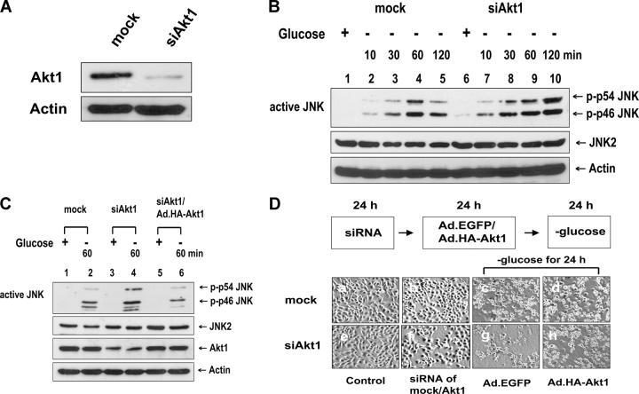
Figure 10.
Role of Akt1 in glucose deprivation–induced JNK activation and morphological damage. (A and B) DU-145 cells were transfected with Akt1 siRNA or mock siRNA and were incubated for 36 h. (A) Akt1 protein expression was assessed by immunoblotting with anti-Akt1 antibody. (B) Cells were exposed to glucose-free medium for various times (10–120 min). Cell lysates were immunoblotted with anti–ACTIVE JNK, anti-JNK2, or antiactin antibody. (C and D) DU-145 cells were transfected with Akt1 or mock siRNA. After 24 of incubation, cells were infected with Ad.EGFP or Ad.HA-Akt1 at an MOI of 10. After 24 h of infection, cells were exposed to glucose-free medium for 60 min (C) or for 24 h (D). (C) Cell lysates were immunoblotted with anti–ACTIVE JNK, anti-JNK2, anti-Akt1, or antiactin antibody. (D) Morphology was evaluated with a phase-contrast microscope.

