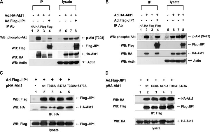
Figure 2.
Phosphorylation of Akt on Thr-308 and Ser-473 and its role in the association of Akt1 with JIP1. (A and B) DU-145 cells were infected with adenoviral vector containing HA-tagged Akt1 (Ad.HA-Akt1) and/or Ad.Flag-JIP1 at an MOI of 10. After 48 h of infection, cells were lysed. Lysates were immunoprecipitated with anti-HA antibody or anti-Flag antibody. Immunoprecipitated proteins and lysates were separated by SDS-PAGE and were immunoblotted with anti–phospho–Thr-308–Akt, anti–phospho–Ser-473–Akt, anti-Flag, anti-HA, or antiactin antibody. (C and D) DU-145 cells were transfected with pHA-Akt1 (wild type), pHA-Akt1 (Thr-308A), pHA-Akt1 (Ser-473A), or pHA-Akt1 (Thr-308A + Ser-473A) plasmids and were infected with Ad.Flag-JIP1 at an MOI of 10. After 48 h of incubation, cells were lysed. Cell lysates were immunoprecipitated with anti-HA antibody (C) or anti-Flag antibody (D) and were immunoblotted with anti-Flag or anti-HA antibody (top). The presence of Flag-JIP1 or HA-Akt1 in the lysates was verified by immunoblotting with anti-Flag antibody or anti-HA antibody, respectively (bottom).

