Figure 5.
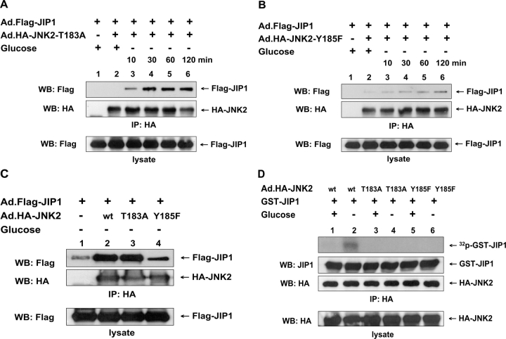
Role of Thr-183 and Tyr-185 residues of JNK2 in glucose deprivation–induced JNK2–JIP1 interaction or JIP1 phosphorylation. DU-145 cells were coinfected with Ad.Flag-JIP1 and adenoviral vector containing HA-tagged wild-type JNK2 (Ad.HA-JNK2-wt), Thr-183A mutant-type JNK2 (Ad.HA-JNK2–Thr-183A), or Y185F mutant-type JNK2 (Ad.HA-JNK2-Y185F) at an MOI of 10. After 48 h of infection, cells were exposed to glucose-free medium for various times (A and B) or for 60 min (C and D) and were lysed. (A–C) Lysates were immunoprecipitated with anti-HA antibody and were immunoblotted with anti-Flag or anti-HA antibody (top). The presence of Flag-JIP1 in the lysates was verified by immunoblotting with anti-Flag antibody (bottom). (D) Cell lysates were immunoprecipitated with anti-HA antibody. To examine which types of JNK2 can phosphorylate JIP1, 0.5 μg GST-JIP1 was incubated with immunoprecipitated HA-JNK2 in kinase buffer containing 100 μCi/ml γ-[32P]ATP at 30°C for 1 h. Phosphorylated proteins were resolved by SDS-PAGE and were analyzed by autoradiography. The presence of GST-JIP1 and HA-JNK2 in the kinase buffer was verified by immunoblotting with anti-JIP1 antibody and anti-HA antibody, respectively (top). The presence of HA-JNK2 in the lysates was verified by immunoblotting with anti-HA antibody (bottom).

