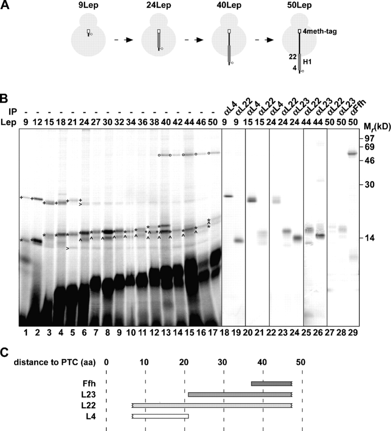
Figure 1.
Progression of nascent Lep through the ribosome. (A) Schematic representation of the 9, 24, 40, and 50Lep constructs with a cross-linking probe at position 3. The transmembrane regions (H1) and methionine tags are presented as thick gray lines and white bars, respectively. (B) In vitro translation of nascent 9–50LepTAG3 constructs was performed in the presence of (Tmd)Phe-tRNAsup. After translation, samples were irradiated with UV light to induce cross-linking, and the ribosome–nascent chain complexes were purified and analyzed by SDS-PAGE. UV-irradiated 9, 15, 24, 44, and 50LepTAG3 were immunoprecipitated with antisera as indicated. (C) Schematic representation of the cross-links observed in B. Numbers indicate the distance in amino acids of the cross-linking probe to the PTC. Images in different panels represent different parts of the gel or different exposure times. +, L4 cross-link; *, L22 cross-link; ^, L23 cross-link; o, Ffh cross-link; >, L4 and L22 cross-link to a truncated translation product.

