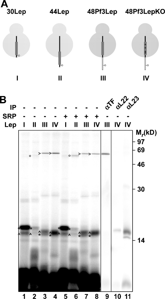
Figure 4.

SRP is not oriented toward the ribosomal exit before it is able to interact with H1. (A) Schematic representation of 30 and 44Lep with a cross-linking probe at position 3 and 48Pf3Lep and 48Pf3LepKO with a cross-linking probe at position seven. H1 and the Pf3 extension are depicted as a thick gray line and a thin white bar, respectively. The four mutations in H1 to obtain the Pf3LepKO construct are the same as in Fig. 3 and are indicated here with four asterisks. (B) The constructs shown in A were translated in vitro with and without the addition of 350 nM of purified SRP. After translation, samples were cross-linked, purified, and immunoprecipitated as described in Fig. 1. Images in different panels represent different parts of the gel or different exposure times. *, L22 cross-link; ^, L23 cross-link; o, Ffh cross-link; >, TF cross-link.
