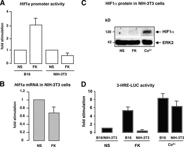
Figure 2.
cAMP induces HIF1α expression in a cell-specific manner. (A) B16 cells and NIH-3T3 cells were transfected with a fragment of the Hif1a gene promoter cloned upstream of the luciferase reporter gene, and were then stimulated (or not) (NS) with forskolin (FK). Luciferase activity was normalized by the β-galactosidase activity and results are represented as the fold stimulation of the cAMP-activated promoter compared with the basal value. (B) Real-time quantitative PCR to detect Hif1a mRNA levels was performed on total RNAs from NIH-3T3 fibroblasts nonstimulated (NS) or treated for 24 h with forskolin (FK). (C) NIH-3T3 extracts were subjected to Western blot analysis to detect HIF1α protein levels after cell stimulation with forskolin (FK) for 24 h, or cobalt (Co2+) for 12 h. ERK2 levels show a control of the gel protein loading. White lines indicate that intervening lanes have been spliced out. (D) 3-HRE-LUC reporter assay on B16 and NIH-3T3 cells nonstimulated (NS) of stimulated with forskolin (FK) or cobalt (Co2+).

