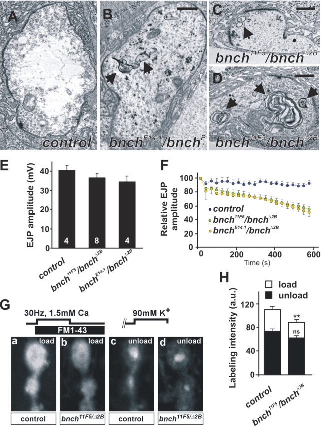
Figure 5.

Abnormal membranous structures and presynaptic endocytic defects at the bnch NMJ. (A–D) Ultrastructure of yw (A, control), yw; bnch E14.1/bnchP (B) and yw; bnch 11F5/bnchΔ2B (C and D) NMJ boutons at rest. Bars represent 0.5 μm. Membranous inclusions in bnch mutant boutons range from smaller (0.2 μm) vesicular structures with a single limiting membrane (C, arrow) to larger multilamellar bodies (0.3–1.2 μm) (B and D, arrows) that are absent in controls (A). (E and F) EJP recordings in 1 mM Ca2+ from muscle 6 in yw (control), yw; bnch 11F5/bnchΔ2B, and bnch 11F5/bnchΔ2B third instar larvae. Quantification of EJP amplitudes shows no significant differences between bnch mutants and controls (E). (F) High frequency (10 Hz) stimulation during 10 min reveals a gradual rundown of the EJP amplitudes in bnch mutants (green and yellow) but not controls (blue). (G and H) FM1-43 dye loading and unloading experiments in yw (control) and yw; bnch 11F5/bnchΔ2B third instar larval NMJs preparations. FM1-43 dye was loaded during 5 min 30 Hz stimulation in 1.5 mM Ca2+ and 5 min rest (G, a and b). Unloading of the RRP of synaptic vesicles was achieved by 90 mM K+ stimulation during 5 min (G, c and d). Note the subtle decrease in FM1-43 uptake in bnch mutants (G, b) compared with controls (G, a). Unloading of the RRP is similar in bnch mutants (G, d) and controls (G, c). Quantification of these results is shown (H).
