Figure 2.
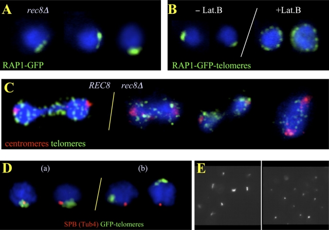
Telomere clustering in rec8Δ meiosis is displaced from the SPB and requires actin polymerization. (A) Rap1-GFP–tagged telomeres (green) in rec8Δ meiocytes display one telomere cluster. (B) rec8Δ meiocyte nuclei before (−Lat B) and after (+Lat B) inhibition of actin polymerization by Lat B treatment, which leads to peripherally dispersed telomeres. (C) Anaphase I figures of wild-type (left) and rec8Δ meiocytes (right) show absence of a telomere cluster. Centromeres (red) lead the anaphase movements, whereas telomeres (green) are seen as numerous spots trailing behind. (D) Spatial relationships of the telomere cluster (green, Rap1-GFP) and SPB (red, Tub4) in rec8Δ meiocytes. Class a nuclei depict abundant patterns of telomere cluster–SPB association in wild-type meiosis. In rec8Δ meiosis, the telomere cluster is often separated from the SPB (class b; Table I). (E) Image field showing tubulin staining (FITC channel, gray scale) of cells from meiotic cultures without (left) and with (right) benomyl and nocodazole treatment. The treated cells display spots as a result of residual SPB staining that is resistant to MT drugs (Hasek et al., 1987; Lillie and Brown, 1998).

