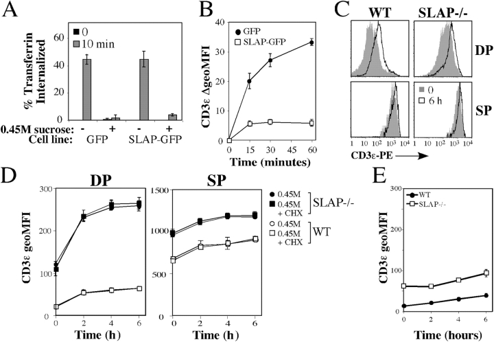
Figure 4.
Increased TCR–CD3 recycling is revealed in hypertonic medium. (A) Uptake of Alexa647-labeled transferrin by Jurkat T cell lines expressing SLAP-GFP or control (GFP) in the presence or absence of hypertonic medium, as assessed by FACS. (B) Expression of CD3ɛ on Jurkat T cell lines expressing SLAP-GFP or control (GFP) incubated in hypertonic medium, as assessed by FACS. Data are presented as the absolute increase in CD3ɛ MFI expression relative to time 0. Data in A and B are the mean of three experiments ± SEM. (C) CD3ɛ expression on WT or SLAP−/− thymocytes incubated in hypertonic medium for 0 or 6 h, as assessed by FACS. Data are representative of three mice per genotype. (D) MFI of CD3ɛ expression on WT or SLAP−/− thymocytes incubated in 0.45 M hypertonic medium for the indicated time in the presence or absence of cycloheximide (CHX). (E) MFI of CD3ɛ expression on WT or SLAP−/− DP thymocytes incubated for the indicated time in cell culture medium only. Data in D and E are the mean of three mice per genotype ± SEM (error bars).

