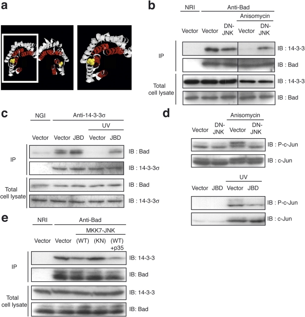
Figure 1.
JNK promotes the dissociation of Bad from 14-3-3 in vivo. (a) The putative JNK phosphorylation site of 14-3-3ζ is adjacent to the ligand-binding groove, which is shown in red. The right panel is a close-up view of the region (white box, left) surrounding the putative phosphorylation site (Ser184), which is shown in yellow. (b) COS-1 cells were transfected with the indicated plasmids and were incubated for 3 h in the absence or presence of 10 μg/ml anisomycin. Cell lysates were subjected to immunoprecipitation (IP) with antibodies to Bad or normal rabbit IgG (NRI). (c) HCT116 cells were transfected with the indicated plasmids and were irradiated by 50 J/m2 UV for 6 h. Cell lysates were subjected to immunoprecipitation with antibodies to 14-3-3σ or normal goat IgG (NGI). (d) Cells were transfected and cultured as described in b and c. Cell lysates were subjected to immunoblot analysis with the indicated antibodies. (e) COS-1 cells were transfected for 18 h with expression vectors for MKK7-JNK and p35 as indicated. Cell lysates were subjected to immunoprecipitation with antibodies to Bad. (b–e) The resulting precipitates were subjected to immunoblot analysis (IB) with the indicated antibodies. The same results were obtained in three independent experiments.

