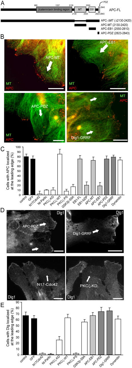
Figure 3.

A Cdc42-PKCζ–dependent, GSK-3β/APC-independent pathway controls Dlg1 localization. (A) APC constructs that were used in this study. Astrocyte monolayers were scratched, and leading edge cells were immediately microinjected with the indicated constructs or incubated in the presence of PKCζ pseudosubstrate (PKCζ-PS; 10 μM for 1 h). Numbers correspond to the amino acid sequences of APC. (B) APC localization visualized with epifluorescence (green, tubulin; red, APC). (C) Percentage of cells with APC clusters at the leading edge. (D) Dlg1 localization visualized with epifluorescence. (B and D) 4 h after wounding, cells were fixed and stained with antibodies recognizing the microinjected constructs. Cells expressing the injected constructs are indicated by an arrow. Similar results were observed 8 h after wounding. Bars, 10 μm. (E) Percentage of cells with Dlg1 recruitment at the leading edge. (C and E) Results are means ± SEM of three independent experiments scoring at least 150 cells.
