Figure 4.

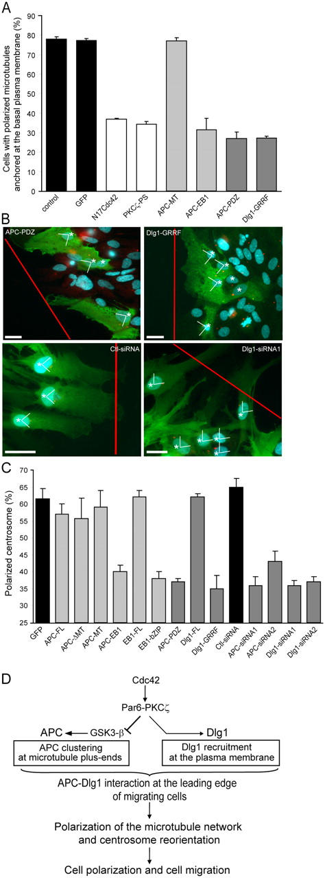
APC–Dlg1 interaction is required for astrocyte polarization. When indicated, cells were nucleofected with pEGFP and siRNA and incubated for 3 d. Monolayers were scratched, microinjected with the indicated constructs, or incubated in the presence of PKCζ pseudosubstrate (PKCζ-PS; 10 μM for 1 h). (A) Polarized microtubule anchoring at the plasma membrane was assessed in astrocytes expressing the indicated constructs. (B) 8 h after wounding, cells were fixed and stained with antibodies recognizing microinjected constructs (green cells expressing the injected constructs are indicated by asterisks), antipericentin (red), and Hoechst (blue). Red lines indicate the directions of the scratch. Bars, 10 μm. (C) Centrosome polarization was assessed in astrocytes expressing the indicated constructs. As described in Materials and methods, 25% of polarized centrosome corresponds to a random orientation. Results are means ± SEM of three independent experiments scoring at least 100 (A) or 300 (C) cells. (D) Schematic diagram showing molecular pathways occurring at the leading edge of migrating astrocytes that control APC and Dlg1 localization and subsequent microtubule polarization and cell migration.
