
Figure 2.
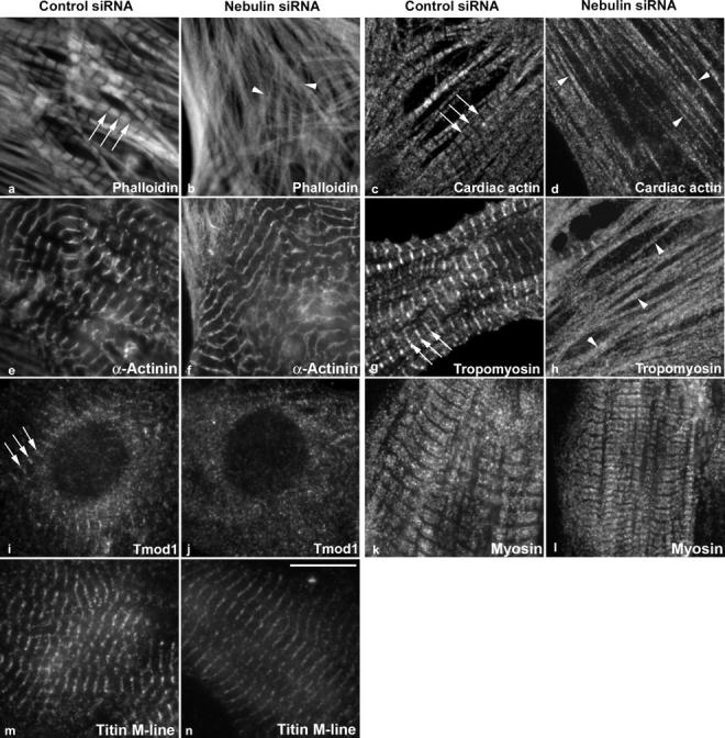
Nebulin is critical for maintaining thin filament lengths from their pointed ends in cardiomyocytes. After 24 h of siRNA treatment, cardiomyocytes were stained with phalloidin to label F-actin (a and b) and with antibodies to cardiac actin (c and d), α-actinin (e and f), tropomyosin (g and h), Tmod1 (i and j), myosin (k and l), and the M-line region of titin (m and n). Note that the staining patterns with both cardiac actin and tropomyosin antibodies are typical in control myocytes. Thin filament components appeared nonstriated in myocytes with reduced nebulin levels (b, d, and h), yet the barbed ends appeared unaffected (f), suggesting that the thin filaments had elongated from their pointed ends across the H zone. Note that myocytes were double stained with phalloidin and for α-actinin (a, e, b, and f). In contrast, the thin filaments in control siRNA–treated myocytes demonstrated regular, striated staining patterns (a, c, and g). Tmod1 staining was absent from the myofibrils in nebulin siRNA–treated myocytes (j). In control and nebulin siRNA–treated myocytes, thick filaments (k and l) and titin (m and n) appeared unaffected 24 h after treatment. Arrows mark striated thin filament components, and arrowheads mark nonstriated thin filament components. Bar, 10 μm.