
Figure 7.
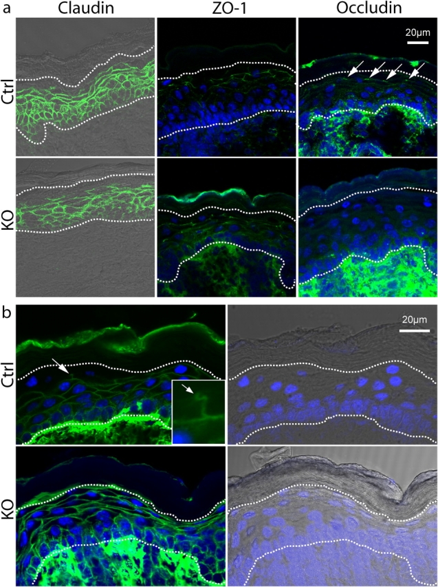
Expression of TJ proteins and TJ permeability assay. (a) Immunofluorescence LSM for claudin-1 (claudin), Zonula occludens 1 (ZO-1), and occludin. For ZO-1 and occludin, the DAPI counterstaining is added. The two dotted lines show the basal membrane (bottom) and the limit SG/SC (top). Claudin staining is localized in the whole epidermis except for the SC and the last layer of SG in both groups. ZO-1 shows a staining in the mid-SG in both groups. In controls, occludin shows a dotted staining, as expected for TJs (arrows), between the second and last layer of the SG. No such staining could be observed in knockouts. (b) TJ permeability assay visualized by LSM. For each genotype, the staining for streptavidin/AlexaFluor488 is shown with DAPI counterstaining, and brightfield/DAPI of the same picture is added for localization. The bottom dotted line indicates the basal membrane, and the top line indicates the limit SG/SC. The top panels show the diffusion of biotin in controls, which is blocked abruptly between the second and last layer of the SG (arrow). The magnification (inset, arrow) indicates the position of TJ. The bottom panel shows the diffusion of biotin in knockout epidermis, which is not blocked by the TJ and, thus, extends up to the SC, where it accumulates.